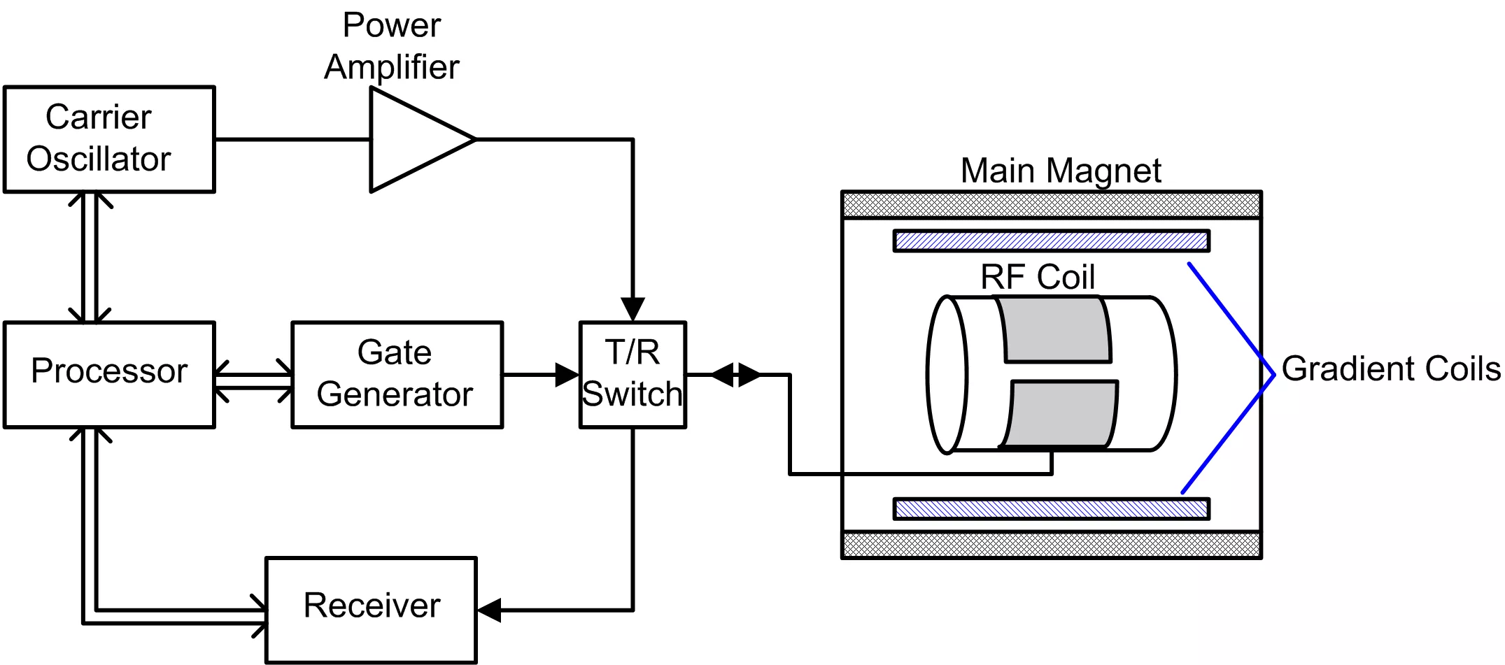
Medical Science
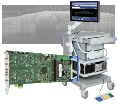
Digitizers are playing an increasing role in medical science particularly when fast electronic signals such as those encountered when using ultrasound, lasers and radiation need to be acquired, analyzed and displayed. The ability of digitizers to convert these types of analog signals into digital information which can then be transferred at high-speeds into computers makes them ideal whenever the information needs to be analyzed and quickly presented. Fast medical imaging is being used to improve diagnosis and help detect disease in the fields of radiology, nuclear medicine, ultrasonography, magnetic resonance imaging (MRI), optical coherence tomography (OCT) and photo-acoustic imaging, dosimetry, positron emission tomography (PET) and other related non-invasive inspection methods.
To cover the broad range and diverse nature of the electronic signals found in medical science Spectrum offers a wide range of digitizers and arbitrary waveform generators. The products are available in a variety of popular standards including PCI, PCIe, PXI and LXI. They offer bandwidths from 50 kHz to 1.5 GHz, sampling rates from 100 KS/s to 5 GS/s, and resolution from 8 up to 16 bits. When large dynamic range and maximum sensitivity is required high-resolution 14 and 16 bit digitizers are available for the capture and analysis of signals that go as high as 250 MHz in frequency. These high-resolution products deliver outstanding signal-to-noise ratio's (up to 72 dB) and spurious free dynamic range (of up to 90 dB) so that small signal variations can be detected and analyzed. They are ideal for use with the sensors used in ultrasound and photo-acoustic systems while the high speed, wide bandwidth digitizers are available to capture the fast pulses (down to the nano and sub-nanosecond ranges) often found in nuclear medicine.
The digitizers are also equipped with ultra-fast trigger circuits, complete with trigger time stamping, so that the dead-time between acquisitions can be extremely small (down to as little as 16 ns). Together with large on-board memories (up to 4 GSamples/card) and advanced streaming and readout modes this makes the digitizers suited to applications where long and complex signals need to be captured and analyzed. Data can be stored in the on-board memory or streamed in FIFO mode over the fast PCIe bus of the digitizer to a PC. By streaming data to a RAID based storage array it's even possible to seamlessly store hours of information. To help with data analysis and data reduction Spectrum's M4i series of digitizers also feature on-board FPGA based processing functions that can be perform on-the-fly Averaging and Peak detection routines.
Each digitizer card can have from one to four channels and up to eight cards can be linked together with Spectrum's StarHub system to create instruments with up to 32 fully synchronous channels, making them perfect for applications where multiple sensors and large sensor arrays are deployed.
Spectrum Product Features
- High Sampling Rates up to 10 GS/s and >1.5 GHz bandwidth
- 12, 14 and 16 bit Resolution
- Fast Trigger and Read-Out Rates
- External Clock and Reference Inputs
- FPGA based Block Average and Block Statisctics (Peak Detect) Options
Matching Card Families
33xx
Family
A/D family
Sample rate
6.40 GS/s - 10 GS/s
Resolution
12 Bit
44xx
Family
A/D family
Sample rate
130 MS/s - 400 MS/s
Resolution
14 Bit 16 Bit
22xx
Family
A/D family
Sample rate
1.25 GS/s - 5 GS/s
Resolution
8 Bit
66xx
Family
D/A family
Sample rate
625 MS/s - 1.25 GS/s
Resolution
16 Bit
Related Documents
Case Study: OCT application for skin cancer diagnosis
The VivoSight® OCT scanner uses the technique of swept-source Optical Coherence Tomography (SS-OCT) for cross-sectional imaging of skin. This is a significant new tool to assist in diagnosis and treatment of skin cancers and other skin conditions.

Case Study: Fast Digitizer from Spectrum enables breakthrough in cell sorting
Cell sorting plays a fundamental role in molecular biology, pathology, immunology and virology research. It requires the ability to rapidly search through and sort out cells based on their unique chemical features and shapes. Conventional methods are limited in uncovering these differences, or are too labor or time intensive, or have to tradeoff between speed and accuracy. The Department of Chemistry at the University of Tokyo has developed an intelligent Image-Activated Cell Sorter (IACS) with an ultra-fast Spectrum Instrumentation digitizer at the heart.
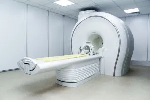
Case Study: Shrunken MRI scanner helps in saving lives of sick babies
MRI scanners are a key diagnostic tool but they are big, very heavy and need liquid helium to cool them. Neoscan Solutions invented an MRI scanner much smaller and lighter which could be placed directly in the children's ward of the hospital, to keep journeys short and to scan sick babies in their sleep.
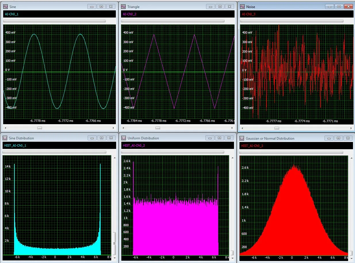
Signal Processing for Digitizers
Modular digitizers allow accurate, high resolution data acquisition that can be quickly transferred to a host computer. Signal processing functions, applied in the digitizer or in the host computer, permit the enhancement of the acquired data or the extraction of extremely useful information from a simple measurement.
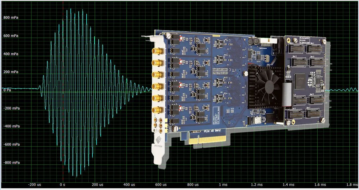
Ultrasonic Applications
The use of Ultrasonic products is increasing as new techniques and improvements in instrument performance constantly expand the range of applications. Spectrum digitizers are ideal tools for making ultrasonic measurements and can play a key role required in the development, testing and operation of these products. Spectrum digitizers and arbitrary waveform generators offer a wide range of bandwidths, sampling rates, and dynamic range to match the broad spectrum of ultrasonic measurement needs
M5i.33xx - Pulse Burst RF Measurements for NMR, MRI and radar
Many RF systems operate using burst modes where radio frequency (RF) signals are transmitted for a short period. Examples of this kind of operation include echo-ranging applications like radar, magnetic resonance imaging (MRI) and relaxation radiation detection in nuclear magnetic resonance (NMR). These applications transmit a burst of relatively high-power RF and then wait for a return echo or a relaxation radiation signal. Measurement of this type of signal requires instruments with wide bandwidth, high sampling rate, long acquisition memory, fast processing, and high-speed data streaming.
Research Papers
Photolysis of Formic Acid
The Department of Chemistry, National Taiwan University, in Taiwan are studying the roaming dynamics and conformational memory in photolysis of formic acid. They use an M4i.4410-x8, 130 MS/s, 16 bit, Digitizer for data acquisition in their Time-resolved Fourier-transform Infrared Emission Spectroscopy system. A research paper on the topic can be found below
Research PaperBiomedical Laser Spectroscopy
At the Norwegian University of Science and Technology they are using an eight channel M2p.5946-x4 80 MS/s, 16 bit, digitizer card as part of their studies into biomedical laser spectroscopy. The development of a Mid-Infrared Laser Spectroscopy for Glucose Sensing is discussed here:
Research PaperOCT - Optical Coherence Tomography
See how fast Spectrum digitizers M2i.2031 and M2i.4022 are used for OCT data acquisition at the University of Sheffield, UK, by clicking here:
Research PaperMPI in Cancer Research
Learn how Japan's Meiji University is using a high-resolution Spectrum digitizer M2i.4031 for the evaluation of magnetic nanoparticle samples in cancer research with time correlation magnetic particle imaging (MPI) by clicking here:
White PaperPhotoacoustic Wavefront Shaping
At London’s St Thomas’ Hospital in the UK, they are working on a high-speed photoacoustic-guided wavefront shaping method, that uses a relatively simple experimental setup, with potential for in vivo applications. Part of the system uses an M4i.4420-x8 250 MS/s, 16-bit Digitizer to acquire ultrasonic signals. The full research paper discussing the experimental setup and results can be found here:
Research PaperUltrasound Endoscope
The College of Biophotonics, South China Normal University, China, is improving the performance of photoacoustic/ultrasound endoscopes. Their research uses an M4i.4420-x8 250 MS/s, 16-bit Digitizer as part of dual-modality endoscope. A research paper shows the potential for using the tens-of-micron-resolved PA/US endoscope for in vivo anatomical imaging in the clinical detection of colorectal diseases.
Research PaperDrug Delivery for Cancer Treatment
The University of Leeds and the Leeds Institute of Medical Research, Leeds, U.K. are studying the use of nanobubbles as a drug delivery agent for cancer treatment. A Spectrum M4i.4420-x8, 250 MS/s, 16-bit Digitizer is used as the data acquisition card that collect acoustic emission signals. A paper discussing the research can be found here:
Research PaperPatienst Safety in MRI Process
At the Physikalisch-Technische Bundesanstalt (PTB) in Germany they are investigating ways to improve the safety for patients with implants who are to be subjected to an MRI process. The PTB is using two M4i.6622-x8 AWG’s and an M4i.4451-x8 digitizer as part of a signal transmission and reception system. A white paper that describes the system and demonstrates how RF induced heating and its mitigation can be achieved is presented here:
White PaperOptoacoustic Image Reconstruction
The University of Bern’s Institute of Applied Physics in Switzerland is testing and developing algorithms used for image reconstruction in optoacoustic imaging applications. Test signals are acquired using an M4i.4420-x8, 250 MS/s, 16 bit, digitizer and a research paper discussing their findings can be downloaded from here:
Research PaperOptoacoustic Microscopy for Dermatologic and Micro-angiography
At Switzerland’s Institute of Pharmacology and Toxicology and Faculty of Medicine, at the University Zurich, they are using a burst-mode laser triggering scheme and an M4i.4420-x8, 250 MS/s, 16 bit, digitizer to perform rapid acquisition functional optoacoustic micro-angiography. A paper discussing the developed system, and how it greatly enhances the performance and usability of optoacoustic microscopy for dermatologic and micro-angiographic studies, can be found here:
Research PaperHandheld Photoacoustic Microscopy
The MOE Key Laboratory of Laser Life Science and Institute of Laser Life Science, at the South China Normal University, in China has developed a Photoacoustic Imaging (PAI) pen that can be handheld (performing forward detection and lateral detection) to extend the application of photoacoustic (PA) microscopy to areas such as the oral cavity, throat, cervix, and abdominal viscera. The experimental setup uses an M4i.4450-x8 500 MS/s, 14 bit, digitizer to acquire the sensor signals. A paper discussing the PAI pen and the test results can be found here:
Research PaperFTIR Spectroscopy for Protein-Membrane Interactions
At the University of Konstanz a fast high resolution Spectrum M2p.5960-x4 digitizer provides nanosecond time-resolution in an FTIR spectroscopy system that’s used to investigate protein-membrane interactions. See the full details here:
Research PaperPhotoacoustic Imaging
Find out how Nanyang Technological University, Singapore, uses a high speed 16 bit Spectrum digitizer M4i.4420-x8 for Photoacoustic Imaging by clicking here:
Research PaperUltrasonic 3D Endoscopic Imaging System
See how the Department of Medical Physics and Biomedical Engineering, at University College London, UK, use an M4i.4420-x8 high-resolution digitizer in a miniature all optical ultrasonic 3D endoscopic imaging system by clicking here:
Research PaperNon-Invasive Cancerous Tissue Treatment
Click below to find out how the School of Electronic and Electrical Engineering, University of Leeds, UK, are using a Spectrum M4i.4420-x8 high-resolution digitizer, plasmonic gold nanorods and high intensity focused ultrasound (HIFU) to improve non-invasive techniques for the treatment of cancerous tissue
Research PaperTherapeutic Ultrasound Brain Deseases Treamtment
At the Queensland Brain Institute, University of Queensland, researchers are using a Spectrum M4i.4421-x8 16 bit digitizer to study ultrasound propagation in materials that are used to modell the human skull as well as investigating therapeutic ultrasound as a potential means to treat deseases of the brain.
Research PaperBreast Ultrasound Scanning System
The Laboratory of Acoustical Waveform Imaging, Department of Imaging Physics, Delft University of Technology, Delft, The Netherlands, is developing the Delft Breast Ultrasound Scanning System (DBUS) as a means for detecting the presence of tumors. Find out how they are using the 14 bit M3i.4142 digitizer by following this link:
Research PaperReal-Time 2D Ultrasound Imaging
A video-rate all-optical ultrasound imaging system, where ultrasound is generated and detected using light, has been demonstrated at the Department of Medical Physics and Biomedical Engineering, University College London, UK. The system uses a Spectrum M4i.4420-x8 high resolution 16 bit digitizer to acquire signals from a broadband photodiode. Details on how the system enables real-time, video-rate 2D ultrasound imaging, at a frame rate of 15 Hz, can be found here:
Research PaperScanning Transmission Electron Microscopy
In Rehovot, Israel at the Weizmann Institute of Science, Department of Chemical and Biological Physics, they are using the M2p.6541-x4 40 MS/s, 16-bit AWG and M2p.5923-x4 20 MS/s, 16-bit Digitizer cards for signal generation and acquisition in Scanning Transmission Electron Microscopy (STEM). A white paper that discusses flexible STEM, with simultaneous phase and depth contrast, is available here
Research PaperFluorescence Imaging Flow Cytometry System
The Department of Chemistry at the University of Tokyo, Japan, is using a 1.25 GS/s M4i.2212-x8 Digitizer as part of a frequency-division-multiplexed fluorescence imaging flow cytometry system. The aim is to allow accurate characterization and classification of a large population of microalgal cells with single-cell resolution for diverse applications such as water treatment, biofuel production, food, and nitrogen-fixing biofertilization. Details of their research can be found here:
Research PaperOptical Resolution Photoacoustic Microscopy
The School of Biomedical Engineering, Tohoku University, Japan is using a 5 GS/s M4i.2230-x8 Digitizer to achieve optical resolution photoacoustic microscopy with sub-micron lateral resolution for visualization of cells and their structures. Details and results of their experimental setup can be found here:
Research PaperFluorescence Lifetime Estimation
At Tokushima University in Japan they are performing fluorescence lifetime estimation by 1-bit photon autocorrelation procedure from time-series data recorded using a high-speed M4i.2220-x8 digitizer. The application details can be found here:
Application DetailsAcoustic-Resolution-based Photoacoustic Microscope (ARPAM)
At the Central South University in Changsha, China, they are using an M4i.2233-x8 high speed digitizer in an improved acoustic-resolution-based photoacoustic microscope (ARPAM) that helps to resolve the conflict between lateral resolution and depth of field. The article is available here:
Research Paper3 Megapixel Ultrasonic Microscope
The University of Helsinki, Finland, has developed a 3 Megapixel Ultrasonic Microscope using a Spectrum M4i.6631-x8 AWG for signal transmission and an M4i.2233-x8 digitizer for data acquisition. IEEE members can download the full article here:
Research PaperLaser-scanning Confocal Fluorescence Microscopy
Laser-scanning confocal fluorescence microscopy is a relatively new and important tool for biomedical research. At the University of Tokyo they are able to demonstrate confocal fluorescence microscopy at a record high frame rate of 16,000 frames/s thanks to new research and the use of a Spectrum M4i.2212-x8 high-speed digitizer. Click below to read the full story.
Research PaperNanopore Analysis
At the School of Chemistry and Chemical Engineering, South China University of Technology, China, they are using a Spectrum M4x.6621-x4 AWG as a precision waveform generator for their research into a Fourier Transform Induced Data Process for Label-free Selective Nanopore Analysis under Sinusoidal Voltage Excitations. A research paper with supplementary documentation showing the experimental setup is available here:
Research PaperHigh-Speed Single-Pixel Imaging
At the Research Institute for Electronic Science, Hokkaido University, in Japan, they can obtain fluorescence images from biological cells using high-speed single-pixel imaging. The method integrates frequency-division multiplexing and time-division multiplexing and applies the combined technique, namely frequency-time-division multiplexing (FTDM), to optical imaging. Signal acquisition includes the use of an M4i.2212-x8 1.25 GS/s, 8-bit PCIe Digitizer, and a white paper on the topic can be found here:
White PaperTarget Tracking
Target tracking has applications in various field, such as biomedical, computer or robotic vision and non-line-of-light motion sensing. At the University of Science and Technology of
China, Hefei, China they have developed a single-pixel imaging technique that can measure zero-order and first-order geometric moments. The information is then used to reconstruct and track the centroid of a fast-moving object in real time. At the core of the system is an M4i.2212-x8 1.25 GS/s, 8-bit PCIe Digitizer that acquires photodiode signals. This allows back-scattered intensity information from the target to be determined. A white paper discussing the research can be found here
Stable Cavitation Activity of circulating Microbubbles
At the School of Sensing Science and Engineering, Shanghai Jiao Tong University, in China they are using a closed-loop feedback controller, based on pulse length (PL), regulation method to improve the temporal stability of stable cavitation (SC) activity. The aim is to achieve controllable and desirable SC activity in target regions for improved therapeutic efficiency and biosafety. A research paper showing the improvements obtained can be found here 7 The setup uses an M4i.4410-x8 130 MS/s, 16-bit PCIe Digitizer to determine the acoustic emission properties of the circulating microbubbles. The digitizer acquires signals produced by a transducer and passes the data at high speed to a computer for subsequent processing.
Reserach PaperDaughter- and Microbubble Studies
Inertial cavitation (IC) of the preformed microbubbles is being investigated for ultrasound imaging and therapeutic applications. However, microbubbles rupture during IC, creating smaller daughter bubbles (DBs), which may cause undesired bioeffects in the target region. The School of Sensing Science and Engineering, Shanghai Jiao Tong University, in China, is researching the properties of the daughter bubbles with the aim of achieving controllable cavitation activity. A research paper on the topic can be found here with the experimental setup using an M4i.4410-x8 130 MS/s, 16-bit Digitizer to acquire the relevant transducer signals
Research PaperHigh-Order Pulse-Echo Ultrasound
The Institute for Biomedical Engineering, Department of Information Technology and Electrical Engineering, ETH Zurich, Switzerland they are researching ways to improve scanning acoustic microscopy by introducing high order reflection pulse-echo (HOPE)-ultrasound. HOPE is a novel method that leverages high order reflections to improve on several aspects of conventional ultrasound imaging. A research paper on the HOPE development, which uses an M4i.4420-x8 250 MS/s, 16-bit Digitizer, to acquire the related transducer signals, can be found here
Research PapersAll-Optical Ultrasound Imaging
All-optical ultrasound (AOUS) imaging, uses light to both generate and detect ultrasound, and is an emerging alternative to conventional electronic ultrasound imaging. At the Department of Medical Physics & Biomedical Engineering, University College London, United Kingdom, they've developed the first AOUS system with a flexible, handheld imaging probe, which represents a critical step towards clinical translation. White papers describing the research, which uses the M4i.4420-x8, 250 MS/s, 16-bit Digitizer for signal acquisition, are available here:
White PaperAgeing Dementia Research
At the Queensland Brain Institute, The University of Queensland, in Australia they are studying possible ways to treat Alzheimer's disease using scanning ultrasound. A paper discussing the research, which uses an M4i.4421-x8 250 MS/s, 16-bit Digitizer for signal acquisition, can be found here:
Research PaperLaser Control for OCT (Optical Coherence Tomography)
The Technische University, at Eindhoven in the Netherlands, has developed a control strategy, that includes the use of an AWG (model M2p.6566-x4 125 MS/s, 16-bit) as the signal source, for a monolithically integrated widely tuneable laser system on InP for optical coherence tomography. A white paper discussing the development is available here:
Reserahc PaperCavity ring-down Spectroscopy
The Information Materials and Intelligent Sensing Laboratory of Anhui Province, Anhui University, China is using an M2p.5941-x4, 80 MS/s, 16-bit Digitizer as part of their research into the development of a CNN-assisted mid-infrared high-sensitivity exhaled ammonia sensor based on cavity ring-down spectroscopy. A reference paper on the work is available below:
Reference Paper3D Photoacoustic Imaging
The School of Engineering Medicine, Beihang University, Beijing, China have developed a technique for compressed single-shot 3D photoacoustic imaging with a single-element transducer. They setup uses an M2p.5961-x4 125 MS/s, 16-bit Digitizer to collect the transducer signals as outlined in the reference paper below
Reference Paper